The Equine-X Blog
Understanding Equine Connective Tissues: Tendons, Ligaments, Bones, and Fascia
Learn about the various types of equine connective tissues including tendons, ligaments, bones, and fascia, their roles, and common issues and challenges that can arise for horses.
Together, we will explore:
what equine connective tissue is and its different categories and sub-categories.
functions of connective tissue, what it is made of, and where it can be found in your horse.
its importance and the benefit it can bring when functioning properly.
what can happen when it is subject to disease and disorders and the impact this can have on your horse, with a nod to the human impact also.
some tips on what you can do to help keep your horse’s connective tissue system healthy.


The horse has 4 kinds of basic connective tissue:
Nervous tissue (as found in the brain, spinal cord, and nerves).
Muscular tissue (as found in skeletal, cardiac, and smooth muscle).
Epithelial tissue (as found in skin, lining of intestines, and lining of the respiratory tract).
Connective tissue (as found between other tissues everywhere in the body).
Connective tissue is our focus here and is the most abundant, widely distributed, and varied type of body tissue. Whilst diverse, they do share multiple structural and functional features which brings them together under the term “connective tissue”.
Equine connective tissue binds the horse’s body together, connects to other tissues and provides support. This means it provides a range of essential and specialist roles in many locations throughout your horse.
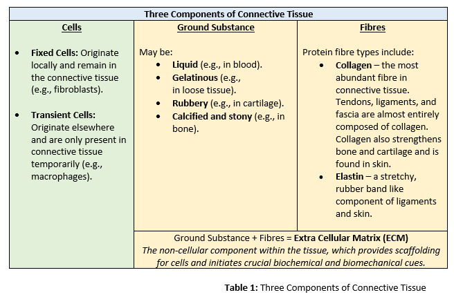
The three components of connective tissue are cells, ground substance and fibres, with the latter two making up what is termed the extra cellular matrix (ECM). Refer to Table 1 for a further overview of these components.
main categories of connective tissue
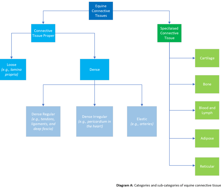
The two main categories of equine connective tissue are Connective Tissue Proper and Specialized Connective Tissue, each of which then have their own sub-categories. Refer to Diagram A to help illustrate.
Connective tissue proper:
a. Loose
b. Dense
Specialized connective tissue:
Cartilage
Bone
Blood and Lymph
Adipose
Reticular
We will look at each of these in more detail below.
EQUINE conective tissue proper
Equine connective tissue proper helps connect tendons, ligaments, muscles, skin, and bone, and is further divided into loose and dense tissues:
Loose
Loose, or areolar, tissue hold organs, anatomic structures, and tissues in place. It is made up of collagen fibres loosely scattered in the ECM. An example of loose connective tissue is lamina propria which is found the beneath the epithelium of mucosal membranes, such as that of the intestinal and respiratory tracts.
Dense
Collagen fibres heavily packed in the ECM either in parallel order (dense regular), or randomly interlaced (dense irregular), or elastic:
Dense Regular (in parallel order), such as that of tendons, ligaments, and deep fascia.
Dense Irregular (randomly interlaced), such as that of the pericardium in the heart.
Elastic, such as that of arteries.
SPECIALIZED EQUINE CONNECTIVE TISSUES
Specialised equine connective tissues are composed of varying specialized cells and ground substance. These diverse but specialized connective tissues include cartilage, bone, blood, lymph, adipose and reticular tissues.
Horse cartilage provides protection, flexibility, cushioning, and support. It is made up of chondroblasts cells which secrete a matrix composed of collagen, elastic fibres and glycosaminoglycans. These cells mature into chondrocytes, which make up the cellular component of cartilage. Cartilage acts as a shock absorber and reduces friction to protect the horse’s joints and bones.
Fibre types found in cartilage are Collagen II (hyaline cartilage), Elastic Fibres (elastic cartilage), and Collagen I (fibrocartilage).
Examples of where cartilage can be found in the horse include but are not limited to:
Knee joints.
Hock joints.
Stifle joints.
Between the vertebrae of the horse’s spine.
Ribs.
Equine bone is specialized for structure, support, and protection of organs. It is made up of osteocytes, which secrete a hard mineralized matrix. The hard exterior surrounds a softer bone marrow interior, which houses stem cells that produce blood cells for the horse. Osteoblasts and osteoclasts are special cells that help horse’s bones grow and develop. Osteoblasts form new bones and add growth to existing bone tissue. Osteoclasts dissolve old and damaged bone tissue so it can be replaced with new, healthier cells created by osteoblasts.
Examples of bones found in the horse’s skeleton include but are not limited to:
Cannon bones.
Skull.
Vertebrae.
Sesamoid bones.
Navicular bone.
Blood and Lymph connect all systems of the horse’s body. Blood transports oxygen, nutrients, and wastes. Blood also helps regulate body temperature and plays a significant role in fluid and electrolyte balance. In addition, it has a protection function through clotting of wounds to prevent blood loss, helping against microorganism invasion (e.g., white blood cells) and disease prevention (e.g., antibodies). Lymph also maintains fluid levels, transports substances, and participates in the immune response.
Adipose tissue is made up of two main cells, white and brown adipocytes. White adipocytes are involved in storage of fat, which tends to be the most well-known aspect of adipose tissue. Brown adipocytes are involved in thermoregulation but are present only in low quantities in horses so are not well documented. An evolving area of research is the role of adipose tissue as an endocrine organ, which will be an interesting area to track for horses with obesity.
Reticular tissue has a branched and mesh-like pattern, often called reticulum, due to the arrangement of reticular fibres (reticulin) organised in delicate networks, which are Type III Collagen Fibrils. Reticular tissue is found in highly cellular locations such as the liver.
Whilst muscle and neural tissue topics may be more familiar to many, connective tissue has the important function of ensuring all the other body systems work in harmony. Meaning that healthy connective tissue allows for more harmony throughout the horse. However, just like the other tissue systems, connective tissue is also subject to disease and disorders, which can then disrupt this overall balance. When this is disrupted, then as a rider you may feel that something isn’t quite right in the horse’s way of going, or if you are an owner then you may hear this type of feedback from your rider, and as a trainer you may visually pick up on cues.
Examples of connective tissue diseases and disorders in the horse include but are not limited to:
Osteoarthritis / Degenerative Joint Disease (DJD).
Sidebone.
Bucked (sore) shins.
Splints.
Chip fragments of bone and cartilage.
Fractures.
Tendon injuries.
Ligament injuries.
Tendon inflammation.
Ligament inflammation.
Degenerative Suspensory Ligament Desmitis / Equine Systemic Proteoglycan Accumulation (ESPA)
Osteochondritis dissecans.
Sacro-iliac disease.
Fascia adhesions.
The impact of which to the horse can range across, depending upon the nature of the issue:
Horse lameness.
Changes in horse behavior.
Pain and welfare concerns.
Reduced range of motion.
The need for corrective shoeing and trimming.
Postural issues.
Gait restrictions and abnormalities.
Compensatory issues.
Poor performance.
Decreased quality of life.
Proprioception challenges.
Whilst the horse impact is of our primary concern, the human impact also includes:
Increased veterinary, farrier, medicine, and care costs.
Loss of training time for horse and rider.
Missed qualifications.
Missed competitions for the horse and rider combination / horse owner.
Confusion and frustration in trying to determine a root cause.
Stress and upset.
Therefore, the benefits of looking after your horse’s connective tissues are as wide reaching as the types of connective tissues themselves. Opportunities to do so for non-inherited connective tissue scenarios include, in no particular order:
Preventing exposure of your horse to toxins.
Providing an appropriate exercise program, with sufficient warm up and cool down periods.
Avoiding long periods of stable confinement.
Giving access to turn out for freedom of movement.
Engaging professionally qualified equine care services for your horse when needed (e.g., equine vet, farrier, horse chiropractor, equine behaviourist).
Using well fitting tack (e.g., saddle, bridle, girth etc.).
Carrying out appropriate stretching techniques.
Keeping your horse well hydrated.
Not using training gadgets that may cause harm.
Continually improving your riding and handling technique with the use of an experienced / qualified coach.
Expanding your knowledge of connective tissues.
Ensuring quality nutrition tailored to the needs of the horse.
Horses in training and competition, as well as leisure horses, invariably face stressors such as increased stable confinement, reduction in pasture turn-out, increased exercise intensity, travel, competition venue stabling and a heightened risk of exposure to pathogens (disease-causing organisms). These stressors tax their connective tissue systems and mean that the modern-day horse can no doubt benefit from additional support.
Feel the difference.
CONNEXION® is formulated to provide nutritional support for equine connective tissues and you can find it online for your horse via the shop page.
Some additional interesting reading and resources you may want to check out are:
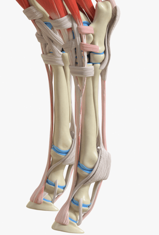
A 3D poster of the hoof palmar systems, showing bones, ligaments and tendons from The Equine Documentalist (T.E.D.): https://www.theequinedocumentalist.com/product-page/ligaments-and-tendons-3d-digit
The role of Adipose Tissue in lipid and energy storage is well documented, however, understanding it as an endocrine organ with a central role in obesity pathology is an emerging area and this British Equine Veterinary Association (BEVA) article may be of interest: Bringing equine adipose tissue into focus - McCullagh - Equine Veterinary Education - Wiley Online Library
A quick read on fascia and equine health: Fascia and Equine Health: An Integral Connection - GRASSROOTS GAZETTE (thegrassrootsgazette.ie)


